
Foot bones anatomy Royalty Free Vector Image VectorStock
The first metatarsal bone leads to the big toe and plays an important role in forward movement. The second, third, and fourth metatarsal bones provide stability to the forefoot. Sesamoid bones: These are two small, oval-shaped bones beneath the first metatarsal on the underside (plantar surface) of the foot. It is embedded in a tendon at the.
.jpg)
Foot Bone Diagram resource Imageshare
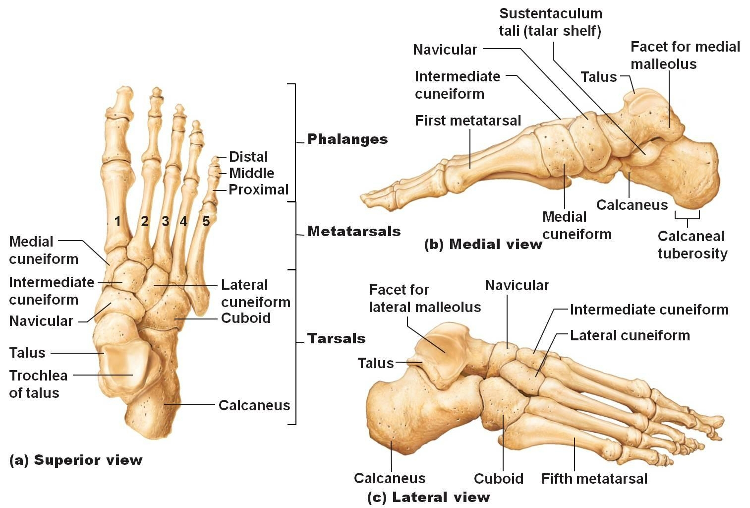
There are 26 bones in the foot, divided into three groups: Seven tarsal bones Five metatarsal bones Fourteen phalanges Tarsals make up a strong weight bearing platform. They are homologous to the carpals in the wrist and are divided into three groups: proximal, intermediate, and distal.

Pin on Anatomy & Physiology
Human body Foot Foot The foot is the lowermost point of the human leg. The foot's shape, along with the body's natural balance-keeping systems, make humans capable of not only walking, but.

Foot bones anatomical vector illustration labeled diagram Human body anatomy, Diagram, Vector
Foot Bones: Forefoot. The forefoot consists of 19 bones; 5 metatarsal bones and 14 phalanges. The big toe has 2 phalanges bones, while the remaining four have 3 phalanges each. The 1st metatarsal is the shortest and thickest of the metatarsals, and it is designed to take up to 40% of your body weight in standing, which rises to 70% when walking.

Anatomy of the Foot and Ankle OrthoPaedia
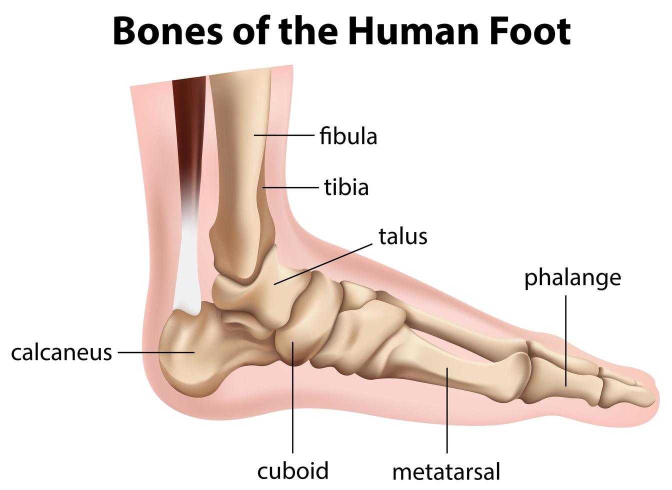
The foot can also be divided up into three regions: (i) Hindfoot - talus and calcaneus; (ii) Midfoot - navicular, cuboid, and cuneiforms; and (iii) Forefoot - metatarsals and phalanges. In this article, we shall look at the anatomy of the bones of the foot - their bony landmarks, articulations, and clinical correlations.
Bones of the human foot diagram 1142236 Vector Art at Vecteezy
The foot itself can be divided into three sections: the hindfoot, midfoot and forefoot and the foot bones can be grouped into three sets: the tarsal bones, the metatarsals and the phalanges .

Ankle and Foot Pain Massage Therapy Connections
The foot is a complex structure comprised of over 26 bones, 30 joints, numerous tendons, ligaments, and muscles responsible for our ability to stand upright, supporting the weight of the entire body and provides the base for the mechanism for bipedal gait. The foot corresponds to the portion of the lower extremity distal to the ankle and divides into hind, mid and forefoot.
Lisfranc Injuries Core EM
Anatomy is a road map. Most structures in the foot are fairly superficial and can be easily palpated. Anatomical structures (tendons, bones, joints, etc) tend to hurt exactly where they are injured or inflamed.

anatomy of the foot Ballet News Straight from the stage bringing you ballet insights
Last updated 2 Nov 2018 The anatomy of the foot The foot contains a lot of moving parts - 26 bones, 33 joints and over 100 ligaments. The foot is divided into three sections - the forefoot, the midfoot and the hindfoot. The forefoot
.jpg?response-content-disposition=attachment)
Foot Bone Diagram resource Imageshare
Use these bones of the foot quizzes to master your identification skills. Overview of the bones of the foot and their divisions into the hindfoot, midfoot and forefoot. With a total of 26 bones in each foot, learning the bony anatomy of the foot is no piece of cake. That is, the memorization aspect.

Foot pain looking up the chain
Foot Anatomy The foot contains 26 bones, 33 joints, and over 100 tendons, muscles, and ligaments. This may sound like overkill for a flat structure that supports your weight, but you may not realize how much work your foot does!

How to have beautiful, healthy feet banish bunions and other abominations! Harrogate Yoga
Tarsal bones - these are the bones closest to the ankle. Each one has a name that translates to describe a little bit about the bone. Talus: "Slope made from rock". Calcaneus: "Heel". Navicular: "Boat Shaped". Cuneiform: "Wedge Shaped". Cuboid: "Cubic in shape". Further along, there are five long bones called.

Foot Anatomy 101 A Quick Lesson From a New Hampshire Podiatrist Nagy Footcare
26 bones 33 joints more than 100 muscles, tendons, and ligaments Bones of the foot The bones in the foot make up nearly 25% of the total bones in the body, and they help the foot.

Foot Bone Anatomy Vector Illustration 539973 Vector Art at Vecteezy
Human body Skeletal System Bones of foot Bones of foot The 26 bones of the foot consist of eight distinct types, including the tarsals, metatarsals, phalanges, cuneiforms, talus,.

Bone Of Left Foot Anatomy Amp Physiology Illustration Human Anatomy Body
The diagram of bones in the ankle and foot is given below: Tarsal Bones The tarsal bones in the foot are located amongst tibia, metatarsal bones, and fibula. There are in all 7 bones, which fall under tarsal bones category. They are: Calcaneus or Calcaneum: To explain the term in layman's language, it is the heel bone in the skeletal system.

The bones in the foot inferior view (Picture illustrated from Thieme... Download Scientific
Summary The foot is an intricate part of the body, consisting of 26 bones, 33 joints, 107 ligaments, and 19 muscles. Scientists group the bones of the foot into the phalanges, tarsal.
